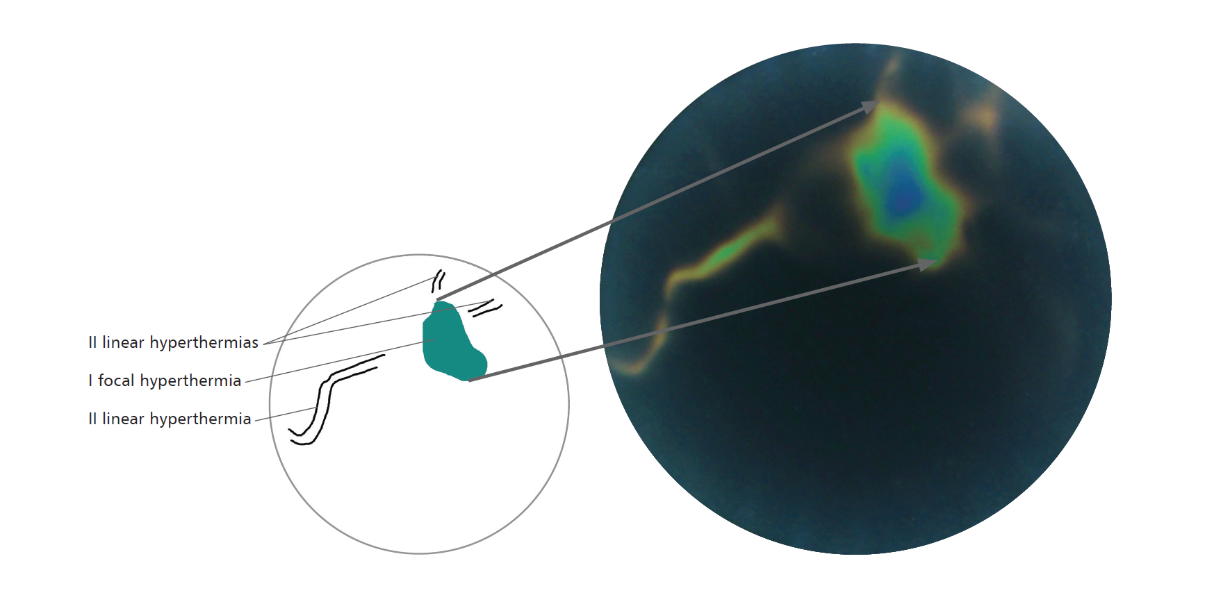
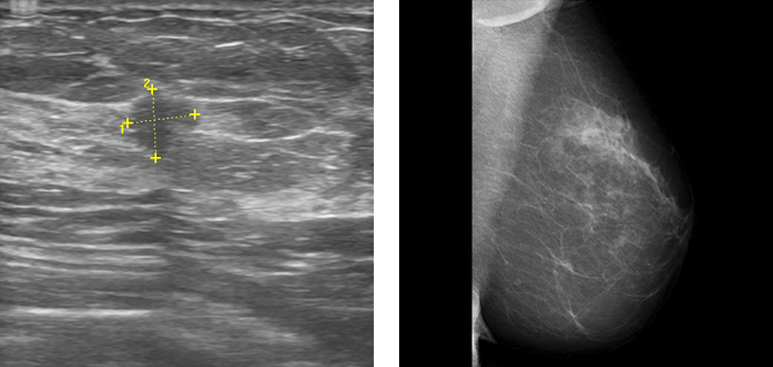
An 89-year-old woman reported to a breast clinic because of a palpable nodule in the left breast. A number of diagnostic examinations were carried out. Thermographic examination preceded by acclimatisation showed a presence of a policyclic hyperthermia focus located in the plane of the upper external quadrant of the left breast. Then mammography was carried out, which confirmed the presence of a spicular densification (with total dimensions, including shoots, of about 40x60 mm, in the upper external quadrant of the left breast (BI-RADS 5)). For the purpose of a thick needle biopsy, USG examination was carried out. In the location of a palpable nodule in the left breast, it showed a policyclic hypoechogenic lesion with dimensions of 25x17 mm (BI-RADS 5). Following regional analgesia, biopsy cores for histopathological examination were collected and the presence of carcinoma mucinosum was confirmed. In this case, correspondence between all three diagnostic methods for detection of breast cancer was demonstrated. In the left breast, an irregular hyperthermia focus (I) corresponding to the verified cancer. Linear hyperthermias in the vicinity (II) corresponding to blood vessels. USG: In the left breast, at 2 o'clock, peripherally irregular hypoechogenic lesion with the dimensions of 25x17 mm; BI-RADS 5. MMG MLO: In the left breast, in the upper external quadrant, policyclic tumour with shoots, with the dimensions of 40x60mm; BI-RADS 5. 


Sign In
Create New Account