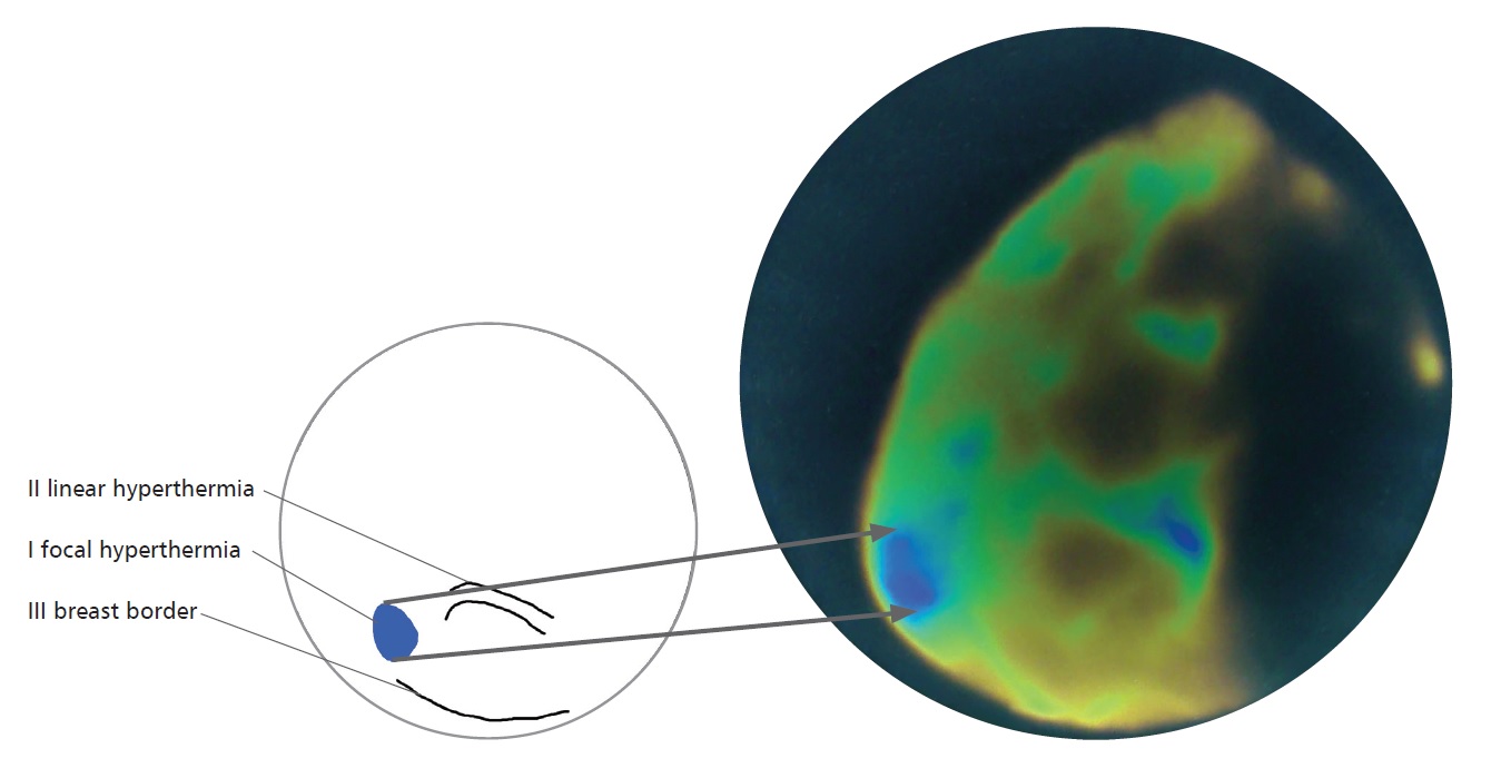
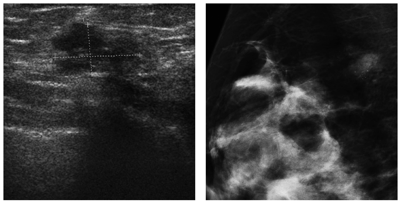
A 37-year old patient came in for a control USG, physical examination was unremarkable, and showed no irregularities. The ultrasound examination revealed dense breast structures. In the right breast, in the 9 o’clock position, an irregular hypoechoic change was visible peripherally with a size of 9x16mm, not well-demarcated, requiring urgent verification by histopathological examination. Prior to the planned biopsy, mammography was recommended to assess both breasts. Mammography, in the case of dense breasts, has limited sensitivity. Mammography revealed the presence of a structural abnormality in the right breast near the spence’s tail, nearly 20mm in size, with poorly saturated microcalcifications. The result was BI-RADS 0 and we recommended to verify the changes under ultrasound. Before the biopsy, a thermographic examination was performed, which revealed the presence of an oval area of hyperthermia in the right breast, in the same quadrant demarcated by the ultrasound examination. Moreover, a linear hyperthermia was visible in the environment suggesting the presence of a blood vessel. A good correlation was found between the available diagnostic tests, which led to the correct diagnosis and possibility of initiating early treatment for breast cancer in this young woman. Ultrasound: In the right breast, at the periphery of the 9 o’clock position, an ill-defined irregularly shaped hypoechoic mass (measuring 16 x 9 mm); BI-RADS 4c**. MMG MLO: In the right breast, in the Tail of Spence, a structure distortion containing low-density microcalcifications (approximately 20 mm in diameter) is visible; BI-RADS 0*,** . *The difference in location is due to the orientation of the breast. **The original description of the examination was preserved; the lesion was histopathologically confirmed as breast cancer.


Sign In
Create New Account