Case Number 2 (104-033)
A 49-year-old woman reported to the oncology center in the town of her residence with palpable lumps in both breasts. In the preceding few years, the patient had not carried out breast examinations.
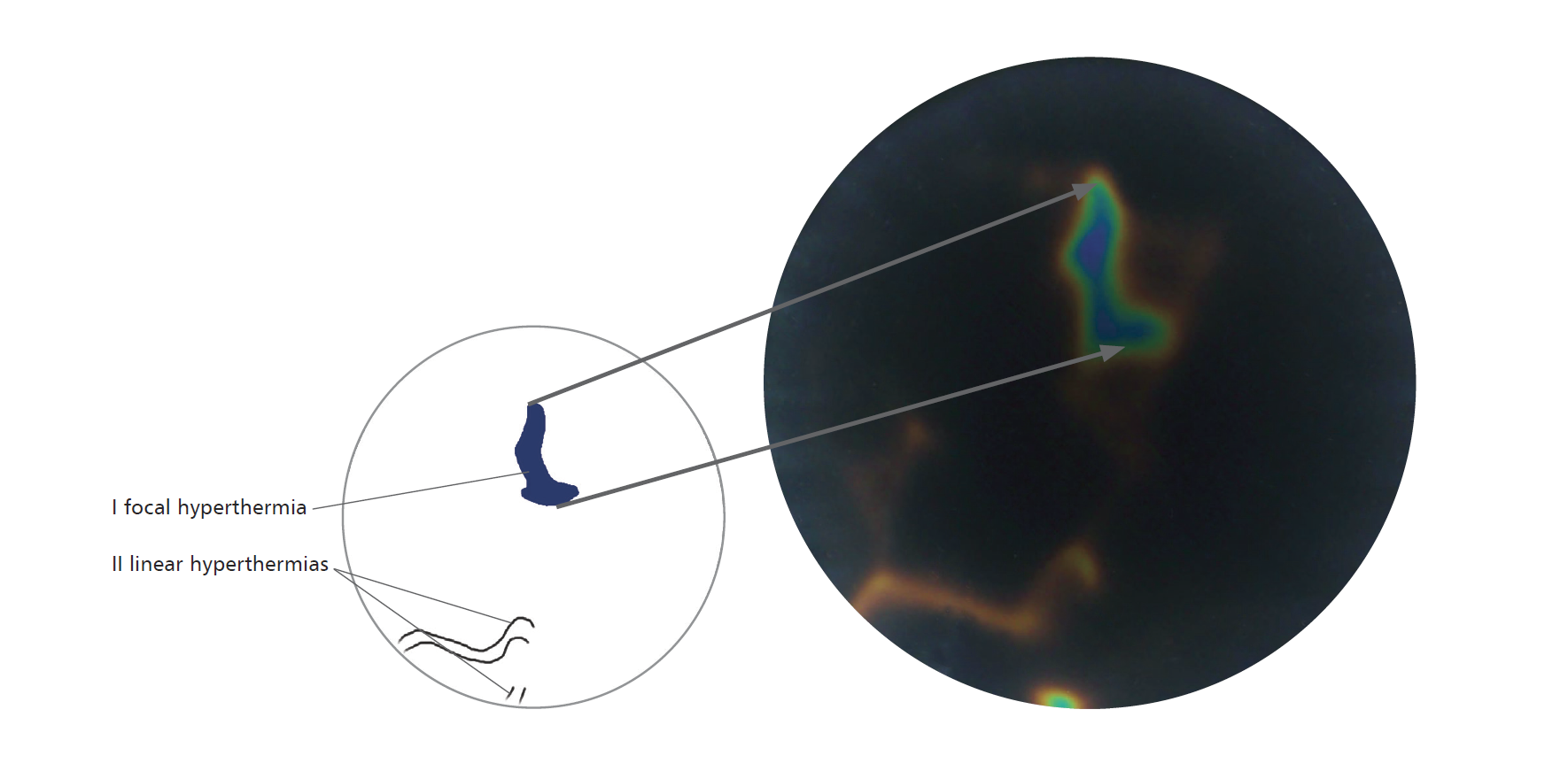
First, thermographic examination with the use of the Braster medical device was proposed, which showed the presence of an irregular hyperthermia focus in the left breast, in the plane of the upper external quadrant.
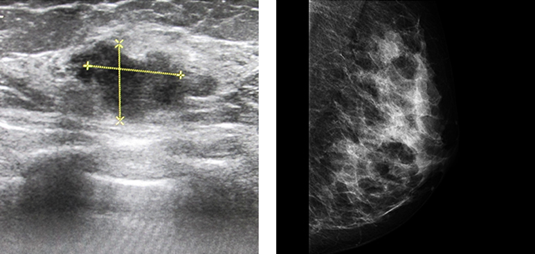
Then, mammography was carried out, which did not show suspicious lesions or micro-calcification focuses (BI-RADS 1).
On the same day, the patient underwent USG examination, which confirmed the presence of a suspicious focal lesion in the left breast. An irregular hypoechogenic lesion with dimensions of 12x16 mm was found at 2 o'clock on the breast's periphery.
Following regional analgesia of the upper external quadrant of the left breast, a thick needle biopsy was carried out with collection of specimens. The histopathological result was acquired: invasive ductal carcinoma.
A significant correlation between the ultrasonographic and thermographic examination of the patient was found. Probably due to the adipose-glandular structure of the breasts, the lesion was not detected in mammography.

In the left breast, an irregular hyperthermia focus (I) corresponded to the verified cancer, on top of that visible linear hyperthermias (II) corresponded to the presence of blood vessels.

USG: In the left breast, at 2 o'clock, peripherally irregular hypoechogenic lesions with the dimensions of 16x12 mm; BI-RADS 4b.
MMG MLO: Mammography does not show suspicious focal lesions or foci of micro-calcifications; BI-RADS 1.


Sign In
Create New Account