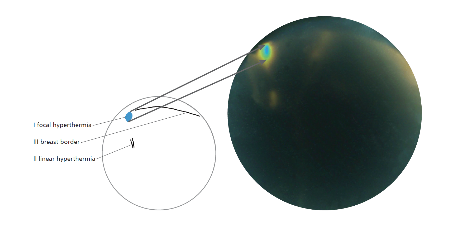
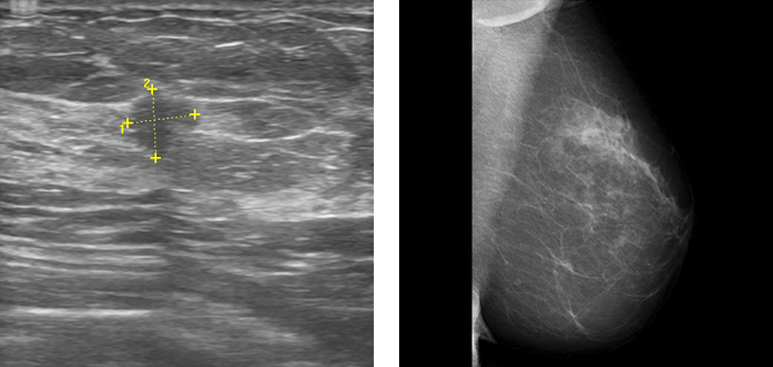
A 60-year-old woman reported to a breast clinic for extended diagnostics after a screening mammography examination had shown pattern distortion at the border of external quadrants of the right breast, BI-RADS 0, for assessment through USG. The surgeon did not detect irregularities through palpation. After appropriate acclimatisation, thermographic examination was carried out with the use of the Braster medical device, which revealed the presence of an oval hyperthermia focus in the right breast, in the plane of the upper external quadrant. Then, a USG examination was carried out, which confirmed the presence of a suspicious lesion in the right breast. A hypoechogenic lesion of an irregular shape with a diameter of 7 mm was found at 11 o'clock at a distance of 5 cm from the nipple (BI-RADS 5). Following regional analgesia of the upper external quadrant of the right breast, the patient underwent a thick needle biopsy and the histopathological result was acquired: invasive ductal carcinoma. In the right breast, oval hyperthermia focus (I) corresponding to the verified cancer, on top of that a visible linear hyperthermia (II) corresponding to the blood vessel. At the top of the image, an outline of the breast border (III). USG: In the right breast, at 11 o'clock, approximately 5 cm from the nipple, a regular hypoechogenic lesion with a diameter of 7 mm; BI-RADS 5. MMG MLO: In the right breast, at the border of external quadrants, distortion of the pattern with a presence of an oval nodule; BI-RADS 0.


Sign In
Create New Account